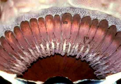
 How does one recognize the vitreous base?
As shown in the photograph above- the vitreous base is pigmented. The gross-dark pigmentation begins on the anterior border of the vitreous base (VB).
Where is the ora serrata located in relation to the vitreous base?
The vitreous base straddles the ora serrata. Notice that the ora serrata is scalloped, a feature that is usually predominant nasally.
Where is the vitreous base wider, temporally or nasally? The ora serrata extends more posteriorly on the temporal side but the vitreous base is actually wider nasally. The scalloped margin of the ora is more prominent nasally. Note the typical cystoid degeneration posterior to the ora serrata.
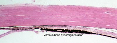 What is the histologic correlate of vitreous base pigmentation?
Above- Pigment epithelial hyperplasia accounts for the pigmentation of the vitreous base. The ora serrata is shown to the left marked by cystoid changes (typical cystoid degeneration).
Below- transillumination highlights the pigmentation of the vitreous base and its anterior and posterior borders as it straddles the ora serrata. This photograph was taken temporally where the ora serrata is less scalloped.

VITREOUS-
What is the volume of the vitreous? The vitreous cavity is simply an expanded extracellular space that normally contains 4.0 ml of clear gelatinous substance that is composed largely of water, hyaluronic acid, and collagen. The vitreous normally contains anteroposterior oriented collagen fibrils and occasional macrophages or hyalocytes. The presence of even small numbers of acute or chronic inflammatory cells within the vitreous is distinctly abnormal. The vitreous has distinct attachments to ocular structures. It is attached most firmly anteriorly in a circumferential band extending from the posterior pars plana to a few millimeters behind the ora serrata in what has been termed the vitreous base.
What is the approximate width of the vitreous base?
The vitreous base is usually 4-5 mm in width. Traction exerted by the vitreous body at the base results in hyperpigmentation of the underlying pigment epithelium and is evident grossly above. The vitreous is also attached to the retina over retinal blood vessels and at the optic disc. These attachments are important to understanding vitreous traction, retinal tears, and retinal detachment, for which vitrectomies are sometimes performed.
<Next topic in Ocular Anatomy>
