ANATOMY OF THE HUMAN EYE

Anatomy of the eye includes lacrimal gland, cornea, conjunctiva, uvea (iris, choroid & ciliary body), lens, blood supply, retina, vitreous & optic-nerve. For ophthalmologists, optometrists, medical, dental, and optometry students, eye-anatomy forms the basis for eye-pathology in diseases: dry eye, retinal detachment, macular degeneration, diabetic retinopathy, eye-trauma etc.

About Mission for Vision

 
Web www.missionforvisionusa.org

Monday, February 13, 2006

Human Lens Histology

The lens is a transparent soft biconvex structure composed of crystallins. The adult lens measures about 9 mm in diameter and is 3.5 mm thick. It is completely enveloped by the thickest basement membrane in the body, the capsule (#1 in photomicrograph ), which is 10-20 µm thick of hyaline material containing type IV collagen. There is a layer of large cuboidal epithelial cells, (the lens epithelium) beneath the anterior capsule (#2 in photomicrograph). 
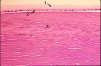
In the center (#3 in photomicrograph) tightly packed cells have lost their nuclei and become packed by special transparent proteins (crystallins) to form so-called lens fibers. New lens cells are added to the margin of the lens throughout life from the lens epithelium, but the cells at the cortex and nucleus (center) of the lens do not undergo turnover or replacement and are therefore the oldest cells in the body of an adult. The lens is avascular and nourished by diffusion from the aqueous and vitreous. The radius of curvature of th
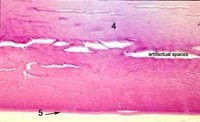
e anterior surface averages 10 mm, but it is subject to marked changes during accommodation. Because the lens nucleus is formed by increasing density of cortical cells, cortex and nucleus are considered together. Both are made up exclusively of cells derived from the lens epithelium. The common designation of “lens fiber” for the cortical cells is a misnomer for the elongated cells of the lens substance. Lens cortical cells are elongated and on cross-section, appear hexagonal in shape. The cortex resembles the cut surface of a honeycomb. In light microscopy, the transition from cortex to nucleus is characterized by less distinct lamellae. The posterior capsule (#5 in the photomicrographs) does not have any epithelium associated with it as the epithelium "migrates" anterior from the lens equator. Below is a photograph at the lens bow. Note the epithelium abruptly stops where the posterior capsule begins. Click to enlarge the photo and then use the back key to return.


Next topic in Eye Pathology

4 Comments:

Blogger Unknown said...

I am extremely impressed. thanks for sharing all information. It is a great post for the people to get the proper information.

Dissertation site

11:23 AM  
Blogger Unknown said...


Furthermore, they can be astonishingly proper fake tag heuer for some an electric with all just like footballers, cricketers, people plus rolex replica sale. Further more they usually are quite a invaluable design for any scholars plus higher education scholars very. Upcoming they can be pretty cute symbols to get tycoons, potential traders plus individuals. In brief, we will express this Armani look at is actually a cure so that you can tag heuer watches. That'sthe reason there are grasped a attentions with poisonous of folks everywhere in the country. In combination with Armani make breitling replica watches, there are plenty of alternative named cartier replica sale out there to choose from sold in the market including Cartier synthetic version different watches, Fossil different watches.


1:04 AM  
Blogger India Web Cares said...

Website Development Company In Delhi : In white label SEO, you use SEO & various other digital marketing solutions for your consumers. In this instance, you don't need to work with any type of new workers or call for brand-new Infographic Submission sites List resources. When you go with white tag services, the other business's name isn't pointed out as well as it helps you expand your network by using even more solutions. Website Designing Company Dwarka

4:06 AM  
Blogger ODESSA said...

This comment has been removed by the author.

5:20 AM  

Post a Comment

<< Home