ANATOMY OF THE HUMAN EYE

Anatomy of the eye includes lacrimal gland, cornea, conjunctiva, uvea (iris, choroid & ciliary body), lens, blood supply, retina, vitreous & optic-nerve. For ophthalmologists, optometrists, medical, dental, and optometry students, eye-anatomy forms the basis for eye-pathology in diseases: dry eye, retinal detachment, macular degeneration, diabetic retinopathy, eye-trauma etc.

About Mission for Vision

 
Web www.missionforvisionusa.org

Wednesday, October 20, 2010

MISSION FOR VISION "SIGHT" MAP

The primary goal of Mission for Vision is to conduct research leading to effective treatments and cures for human eye diseases. The top priority is funding basic science or clinically relevant projects that search for understanding the causes of human-eye-disease. Mission for Vision also seeks to provide information related to eye disease for the public. Below are some of our informative sites.
Tutorials and Study Guides (free)
Anatomy of the Eye
Ocular Pathology Tutorial
Study Guide for Anatomy of the Eye, Orbit, Related Neuroanatomy-http://www.medrounds.org/
Ophthalmology Resident Manual (
http://www.medrounds.org/)
Pathology Board Review Articles

Tuesday, October 19, 2010

EYE ANATOMY- HUMAN

There are several options for finding information. For UCLA students on their one week required clerkship in Ophthalmology or if you are interested in learning anatomy of the eye in detail just follow the tutorial one page at a time by clicking on the "NEXT" button below after reading this page. You may continue through the tutorial by finding the "Next" button or a "Link" to queries that the page presented. The tutorial is written so you can test your knowledge as you proceed. Be sure to read this entire page to find the "NEXT" button. If you click on sublinks on other pages you may find your way to the vasculature of the eye site prematurely which is the end point for the UCLA tutorial. So for UCLA Students be sure to return to the page prior to the sublink, go to the bottom and click the "NEXT" button.
For those that just want to survey the subject, click on the links below one at time and return to this page each time. To link quickly to a particular topic use the drop down list above or use Google to search this site.

Topics in the Tutorial of Ocular Anatomy


To access these topics click on the linked subject, use the drop down list or simply read each topic and link to the answers or next button to move through the tutorial. We begin with an overview.

A horizontally cross sectioned human eye is shown here. CLICK ON THE PHOTO TO ENLARGE IT AND IDENTIFY THE NUMBERED STRUCTURES. Check your answers below.
The key for the various tissues:
1. Cornea- composed of 5 layers, epithelium, Bowman's layer, stroma (the thickest portion), Descemet's membrane and the endothelium.
2. Lens- composed of an anterior lens capsule, epithelium, cortex nucleus and posterior capsule.
3. Iris- the white stroma is sandwiched between the light brown anterior border layer and the dark brown posterior pigmented layers
4. Sclera- the white tunic protects the inner structures. Thinnest over the insertion of the rectus muscles, the sclera is prone to rupture at this site from trauma.
5. Macula (fovea just below the number). This is the area of central and color vision. Acuity is greatest in this region.
6. Optic Nerve Head (notice the adjacent retinal blood vessels).
7. Retinal vessels supply most of the retina. Choroidal vessels supply the photoreceptors and the underlying choroid.
8. Vortex Veins drain the choroid and as indicated, the coalescence of orange vessels that form a whorled appearance.
More detailed information can be found in our links.

UCLA students Click on NEXT Topic in Ocular Anatomy

Monday, October 18, 2010

ANTERIOR SEGMENT OF THE EYE


Photograph of a human eye that has been bisected in the coronal plane to show the view of the anterior segment from a posterior perspective (as though you are looking from the retina). The crystalline lens is suspended by delicate fibers called the zonule. The ciliary body (CB) is composed of about 72 processes that make up the pars plicata and a flat area called the pars plana. The ora serrata (ora) is the place where the retina joins the ciliary body.

Below is a cross section of an eye with the various structures numbered. Click on the photograph to get higher magnification and then after you have identified the structure, verify by checking the key below.


1. Epithelium (cornea)
2. Stroma (cornea)
3. Descemet's membrane and endothelium (cornea)
4. Anterior chamber
5. Iris
6. Lens
7. ciliary body
8. sclera


NEXT

Sunday, October 17, 2010

Conjunctiva

Conjunctiva (named because it conjoins the eyeball to the lids) is a thin, transparent mucous membrane that lines the posterior surface of the lids, and is then reflected forwards on the eye. Conjunctiva is continuous anteriorly with the epithelium of the cornea. Recessed in the eyelids, the conjunctiva forms a cul de sac, which is open in front at the palpebral fissure, and only closed when the eyes are shut. Although all parts of the conjunctiva are continuous each has been given its own name to emphasize anatomic differences. The palpebral portion lines the eyelids; that portion joining the eyeball is the bulbar conjunctiva and that forming the conjunctival sac and reflecting on the eye is called the fornix. The palpebral conjunctiva is subdivided into marginal, tarsal, and orbital zones. The marginal zone transitions between skin and conjunctiva and shows minimal keratinization. The tarsal conjunctiva is a fairly flat layer. The orbital zone shows more numerous Goblet cells.
The regional variation of the conjunctiva start with an overview in a photograph in which the patient is looking up. The limbus (1) is the junction of the conjunctiva and cornea. The bulbar conjunctiva (2) covers the eyeball and extends into the recess created by forniceal conjunctiva (3). The tarsal conjunctiva (4) covers the tarsus. The marginal conjunctiva (6) is at the eyelid margin where the epithelium will begin to be keratinized. The punctum (5) is also shown.
A sagittal or vertical section of both eyelids and the eye are shown to the left. The cornea (1) and lens (2) provide orientation.
The fornix (3) has more redundant conjunctiva. The marginal conjunctiva(4) and tarsal conjunctiva (6) are indicated. The palpebral portion of the lacrimal gland (5) is also shown in this photograph. The composition of each of these regions varies in the Goblet cell density within the epithelium. In addition, note the greater length of the tarsus and higher number of Meibomian glands in the upper eyelid compared to the lower eyelid. This has implications for the origin of sebaceous carcinoma. This photograph also give a view of the cross section of the eyelid and the alert student will notice the skin externally and orbicularis muscle.

The histology of the conjunctiva varies according to its topographic location. The bulbar conjunctiva is relatively less undulating and contains fewer Goblet cells. In this electronmicrograph the microvilli cover the superficial layer of the epithelium (arrow 1) and the mucin granules (2) of the Goblet cell are captured in a plane lacking contiguity with the surface. The Goblet cells produce gel forming mucins called MUC5AC that may be critical to providing lubrication to the ocular surface.

The tarsal conjunctiva shows a stratified squamous epithelium (1) that has few Goblet cells (none seen here) overlying a very dense fibrous stroma, tarsus (2). Meibomian glands are embedded in the tarsus.

This photomicrograph from the fornix shows numerous goblet cells (7) in infoldings of conjunctiva that form the pseudoglands of Henle (8). Striated muscle (9) is seen beneath the substantia propria.

The puncta open on to the marginal portion of the conjunctiva, and through them the conjunctival sac becomes directly continuous with the inferior meatus of the nose via the lacrimal passages. The conjunctiva contains specialized folds or bumps called the plica semilunaris (arrow 10 in the clinical figure) and caruncle (arrow 11). The plica semilunaris lining contains Goblet cells while the caruncle may have hair, sebaceous glands emanating from the surface. The histologic appearance of the caruncle is shown. Transitioning to a stratified squamous epithelium with few goblet cells (area encompassed by arrows at 11), the caruncle may show focal surface keratinization (arrow 14). The caruncle contains hair follicles (arrow 12), sebaceous glands (arrow 13) and adipose tissue (arrow 15).

NEXT Topic in Ocular Anatomy

Conjunctiva

Questions:
1. What structures does the conjunctiva cover?
2. What type of epithelium makes up the conjunctiva?
3. How does the type of epithelium vary according to topographic location?
4. Is the conjunctiva normally lined by keratin in the outer layers?
5. What specialized cells are contained in the conjunctiva?
5. What do Goblet cells secrete?
6. Based on the anatomy what types of cancers would you expect to be found in the conjunctiva?
7. In the photo below identify the various topographical areas of conjunctiva.

Click on the photo below to enlarge and identify the structures that are numbered. This is a sagittal section of the orbit to include the eye with the upper and lower eyelid. (Each photo has its own number system that point to different structures so you must check each photo separately. )
1. How does the upper lid tarsus differ from the lower lid tarsus?
2. Which eyelid has more meibomian glands in the figure?


How does the epithelium in this view of tarsal conjunctiva differ from that of closer to the fornix? Specifically what cells are less numerous here?
How is the epithelium different?


The cell (#2) in the electron micrograph corresponds to which cell in the photo above?

Click here to link to the answers for all photos.

Saturday, October 16, 2010

Cornea

Here are some questions to help you study.
1. What is the structure of the cornea? Name the layers.
2. Name each of the cell types within the corneal epithelium.
3. What is the composition of Bowman's layer, Descemet's membrane, and the stroma?
4. Why is the cornea clear?
5. What causes the cornea to become cloudy?
6. What is the function of the corneal endothelium?
7. How thick is the central cornea?
8. How thick is the peripheral cornea?
9. IDENTIFY THE LAYERS OF THE CORNEA THAT ARE NUMBERED.


The answers are provided in the link.

Friday, October 15, 2010

Cornea Histology


What are the dimensions of the cornea? The cornea is the transparent disk-like anterior portion of the eye, and in the adult typically measures 10.5 mm from top to bottom and 11.5 mm from side to side. Centrally the cornea is about 500 microns (515-539) in thickness and peripherally it is about 650 microns thick. The cornea is more curved than the eye and protrudes anteriorly. The cornea has five layers.


Corneal epithelium (#1 in Figure)
How many layers does the corneal epithelium have? The non-keratinizing squamous epithelium with a basal cell layer gives rise to five to six superficial layers with a total thickness of about 50 µm.
What nerves innervate the cornea and how anterior do they course?
Numerous free nerve endings terminate in this epithelium and are the afferent part of the blink (ciliary) reflex, which is mediated mainly through the sensory part of the fifth cranial nerve (V1). The nerves generally arise from the long posterior ciliary nerves although some come from conjunctival and episcleral origins. At the ora serrata the nerves branch to distribute all about the limbus. Most studies indicate that the nerves are mainly present in the anterior two-thirds of the cornea and that Descemet's membrane is bereft of innervation. The nerves that enter at the limbus, lose their perineuria as well as myelin sheaths, and advance more radially toward the cornea center in the stroma. The sub-epithelial or sub-basal plexus is particularly dense with innervation. The nerve send branches at 90 degree angles toward the surface and penetrate Bowman's layer. The nerves completely lose their myelin sheaths as they penetrate Bowman's layer. The nerves penetrate up to 2 cell layers from the surface (near the superficial cells). One can imagine that an oblique flap made at 100 microns in the depth of the cornea (as in LASIK) would transect the corneal nerves in the field!
Name the cell layers of the corneal epithelium? The basal layer is composed of a single layer of cuboidal to columnar cells (~ 18 μ in height). Above the basal cell layer are 2 layers of wing cells (shaped like a wing) . Beyond this layer two superficial cells become more flattened and more dense with some loss of cell organelles.
What is the nature of the specialized corneal surface? The surface or superficial cells have glycogen granules as well as a thick outer membrane (100 angstroms) that is PAS positive. The surface glycocalyx is composed of transmembrane mucins, MUC1 and MUC16 that emanate from the surface. (
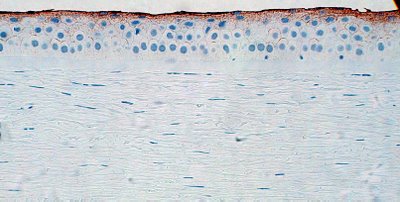
Ref and those within) In the image below the brown areas represent immunohistochemical staining to the superficial corneal epithelium (normally about 2 layers) by antibodies to the 0-linked mucin called MUC 16. The surface cells are covered by a microvillous/microplical surface, interconnected undulations that measure about .5 microns in thickness and height and 1-3 microns in length, not visible in this photograph.
Bowman’s layer (#2 in Figure)
What is the composition of Bowman's layer?
Bowman's layer is composed of fine collagen fibrils and is about 10 µm thick. The layer is acellular. Bowman's layer is limited anteriorly by the basement membrane of the corneal epithelium. The layer is composed of collagen fibrils arranged in random distribution. Where Bowman's layer joins underlying lamellar stroma, there is a transition zone from collagen fibrils that are obliquely arranged to collagenous lamellae of the superficial corneal stroma. Bowman's layer is rarely seen at the slit lamp and only by the most experienced observers. However with appropriate lighting Bowman's layer is quite visible under the dissecting microscope (arrow 2 in the Figure below). Number 3 is on the corneal stroma, 4 is in the anterior chamber and arrow 5 points to the iris.

Corneal stroma (#3 in Figure)
What is the composition of the corneal stroma? The main layer of the cornea is composed of 60-70 successive layers (lamellae of obliquely oriented tightly bound, collagen fibers (corneal lamellae) embedded in an extracellular matrix composed mainly of sulfated glycosaminoglycans. To provide maximum mechanical strength the direction of the collagen fibers differs in each layer. Between the lamellae are sparse fibrocytes (keratocytes).
Does the normal cornea have blood vessels? There are no blood vessels in the cornea; the regular parallel arrangement of the collagen and the paucity of cells render the cornea translucent and allow it to transmit light.
Why is the cornea clear? The cornea is composed of collagen. Collagen in the body is usually opaque. Because the corneal fibrils form a sort of 3 dimensional array of diffraction gratings lamellae, scattered light is eliminated by destructive interference. The fibrils however must be separated from each other by less than ½ of a wavelength of light to remain transparent. Hence the irregularity of Bowman’s layer can be compensated by this arrangement. Corneal edema renders the cornea cloudy because the fibrils become more separated.
Descemet’s membrane (#4 in Figure) which closely resembles the lens capsule, is a true PAS-positive membrane. Produced by the endothelium, the membrane is thin in infancy, increases in thickness to ~ 5 μ in childhood, and then to ~ 8 to 10 μ adulthood.
Corneal endothelium (#5 in Figure)
What is the structure of the corneal endothelium? The endothelial cells lie on the posterior surface of the cornea and form the anterior boundary of the anterior aqueous chamber as a single layer (~ 5 to 6 μ thick) of flattened hexagonally arranged cells.
What is the function of the corneal endothelium? Endothelial cells possess numerous mitochondria, are linked together by both desmosomal and occluding junctions, and pump fluid from the corneal stroma. This thereby prevents excessive hydration of the extracellular matrix, which would result in opacification of the cornea as the separation between lamellae exceeds ½ of a wavelength.

NEXT Topic in Ocular Anatomy


Thursday, October 14, 2010

What is the thickness of the sclera?

The sclera is the white tunic that covers and protects the eye. At the insertion of the rectus muscles (posteriorly) it is thinnest 0.3mm. At the equator it measure 0.4-0.5 mm and at the posterior pole in measures 1.0 mm. The thickness of the sclera is relevant to areas prone to rupture. Blunt trauma most frequently results in rupture of the eye at the thinnest site, behind the insertions of the rectus muscles.

At the entry site of the optic nerve, the sclera is perforated in a sieve like structure to admit optic nerve bundles, axons of ganglion cells. This sieve of sclera is called the lamina cribosa. In addition there are other channels through the sclera called emissaria. Posteriorly there are aperatures around the optic nerve through which the long and short posterior ciliary arteries and nerves pass. About 4 mm posterior to the equator apertures permit the passage of the vortex veins. Anteriorly, anterior ciliary vessels, branches of vessels to the rectus muscle, and nerves pass.

Recurrent nerve loop of Axenfeld- Described in 1895 by T. Axenfeld was a nerve that makes a loop in the sclera (arrow 1) anteriorly. The nerves travel through the sclera usually originating from the long ciliary nerves (arrow 2) and some may approach the surface of the sclera about 1-6 mm from the limbus (arrow 1). The nerves bend 180 degrees creating a mushroomed loop at the surface (arrow 3). The nerve loops have been described to produce symptoms of irritation and tenderness. A study by Stevenson showed that about 12% of eyes have nerve loops and that the nerve loops are bilateral in 1.0%. Topographically, nerve loops were not found in the horizontal plane anterior to the medial or lateral rectus muscles. Most (70%) are found inferiorly. 62% are found anteriorly to the vertical rectus muscle insertions (44% inferiorly + 18% superiorly). 38% are found in the quadrants particularly in the inferonasal quadrant (18%). The nerves are accompanied by pigment and may produce an elevated nodule on the sclera surrounded by a ring of pigment. The ignorant well meaning resident may foolishly attempt biopsy, exquisitely painful for the patient. For more information click this link.

The sclera is composed of fibrous tissue ranging in thick from 10-140 microns. The sclera is rich in elastic fibers and contains smooth muscle.


References:
Axenfeld, T Heidelberg, Kongressber. 1895 p.122
Stevenson, TC, Intrascleral nerve loops, AJO 1963, 55:935.