General features and boundaries of the ciliary body. The ciliary body is shaped like a triangle with the base at the iris root anteriorly and its apex at the ora serrata posteriorly.
What is the pars plicata? The pars plicata contains about 70 ciliary processes, which are ridges or folds 2 mm long, each with a core of stroma and blood vessels and covered by two layers of columnar epithelium.
What is the pars plana? The pars plana is a posterior flat area 4 mm long. Its stroma is continuous with the choroid, whereas the outer pigmented epithelium of the ciliary body is continuous with that of the retina at the ora serrata.
What is the internal lining of the ciliary body? The ciliary body faces the anterior chamber, posterior chamber, and vitreous cavity and is lined by two neuroepithelial layers, a non-pigmented layer internally and a pigmented layer externally. The outer layer of epithelium is pigmented, but the inner layer, which is in contact with the aqueous is non-pigmented. The ultrastructural features of these non-pigmented cells are those of ion-pumping cells; they secrete the aqueous.
What is the structure and function of ciliary muscle? Beneath the epithelium are ciliary muscles, smooth muscles. Contraction of the ciliary muscle lessens tension on the suspensory fibers of the lens and allows the lens to assume a more spherical shape, enhancing the power of the lens during accomodation. The lens also moves anteriorly during this action, which also aids accomodation. The types of ciliary muscle are important for the resident in ophthalmology. There are actually 3 distinct anatomic types of ciliary muscle, longitudinal fibers, radial fibers and circular fibers. The architecture of the fibers have some general principles. All 3 are attached anteriorly at the ciliary tendon (scleral spur and surrounding soft tissue) as Y shaped extensions with the Y inserting anteriorly into the tendon. The longitudinal fibers merge posteriorly and join the sclera as star shaped attachments referred to as episcleral stars. The middle layer or radial fibers run obliquely to merge and attach to the ciliary processes. The fibers of the inner circular layer have very wide Y shaped insertions to relatively distant parts of the ciliary tendon and additional anterior fiber attachments to the iris. These fibers then attach to the sclera but more anteriorly than the longitudinal muscle. The obtuse Y angle of the circular muscle and short distance extending posteriorly to the sclera permit the fibers to run in a largely circular direction. The variation in the type of ciliary muscle that is torn distinguishes traumatic angle recession from cyclodialysis (
see link).
Click on the photo or right click for higher magnification. 1- A faint zonular fiber is seen traversing along the ciliary pr
ocess posteriorly toward its insertion in the pars plana.
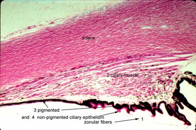
The histologic sections of ciliary muscle show pink smooth muscle bundles (number 2); some are longitudinally oriented toward the scleral side and others are more circular toward the vitreous. The ciliary muscle is responsible for accommodation. The ciliary epithelium is composed of the inner non-pigmented layer (barely visible here at arrow 4) and outer pigmented layer (arrows 3) . The aqueous is made by the non-pigmented epithelium. The beginning of the pars plicata shows the undulations in the ciliary epithelium. Sclera as marked forms the outer tunic.
What is the blood vessel in the center of the ciliary body called? Also seen in this photograph is the prominent central blood vessel in the ciliary body (just to the left of #2 label for ciliary muscle). This is part of the vascular system that is the
major arterial circle, which of course supplies the iris. Tears in this vessel is frequently the culprit in traumatic hyphema. Its position near the border of the longitudinal ciliary muscle explains the predisposition to traumatic injury in
acute angle recession. The circular of the ciliary body and iris is complex and involves
3 anastomotic circles.
What is Axenfeld's nerve loop? 
The intrascleral ciliary nerve loop of Axenfeld is an extension of the posterior ciliary nerve, which makes a loop (arrow 3) through the sclera (arrow 5) in the region of the anterior ciliary arteries or between the rectus muscle insertions. In the figure below the nerve is posterior to the junction of the ciliary muscle at the pars plana. The ciliary epithelium is evident (arrow 1) and so are the lens zonular fibers (arrow 2) as they sweep back along the pars plana. The nerve loop is smooth and dome shaped measuring 1-2 mm in size. The loop may appear cystic and or pigmented leading to a surgical misadventure. The loops are present in about 12% of human eyes. Virtually all are associated with blood vessels and all are pigmented. The loop may protrude to cause symptoms but most importantly, Axenfeld's nerve loop is in the differential of a fixed pigmented epibulbar lesion. It is frequently misdiagnosed as a nevus or melanoma. Manipulation of the lesion elicits pain from the subject as the nerve is present at the external surface (arrow 4). This is the sign that should make one reconsider biopsy! Infrequently, the nerve loops may be multiple in the same eye (0.5%) or bilateral (1%) . For other photographs and additional information about the topography of Axenfeld's nerve loop click
this link.
. <
Next Topic Ocular Anatomy>
