The retina is composed of pigment epithelial cells, photoreceptor cells, retinal support cells and nerve cells that are organized into simple but distinct layers starting with photoreceptors and moving inward to the layer that includes modulating interneurons, and synapsing finally on optic nerve cells (the ganglion cells). The vernacular divides this simplicity into a complex anatomic appearance. Click to enlarge the photomicrograph to the left.
Internal li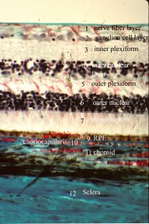 miting lamina
, basement membrane at the interface between the vitreous and the retina.
Nerve fiber layer, axons of the ganglion cells en route to the CNS
Ganglion cell layer, cell bodies of the ganglion cells (large nuclei and nucleoli;
Inner plexiform layer, cell processes and synapses of bipolar cells, amacrine cells, interplexiform cells and ganglion cells
Inner nuclear layer, nuclei and cells bodies of bipolar cells, horizontal cells, interplexiform cells and amacrine cells, as well as the nuclei of the Muller’s cells
Outer plexiform layer, synaptic connections between photoreceptor cells, bipolar neurons and horizontal cells;
Outer nuclear layer nuclei and cell bodies of the photoreceptor cells, 8-9 nine rows;
External limiting membrane (Photo the line between 6-7) adherent junctions between Muller’s cells;
Photoreceptors (photo #7), inner and outer segments of rods and cones
Retinal pigment epithelium (9 (RPE)) sits immediately on
Bruch's membrane (8).
Above the RPE or on the inner or internal side lies the photoreceptor layer of the retina that has the rods and cones. The RPE are supportive cells and have macrophage function. The photoreceptors called rods are about 2 µm thick and average of 50 µm in length, possessing a roughly cylindrical outer segment. Cones are shorter and thicker, 3-5 µm thick x 40 µm long; their outer segment is conical in shape. The tips of these rods and cones are embedded in the microvilli of RPE, which phagocytose the old photoreceptor disks. Cones, perceive color, are concentrated in the macula and particularly the fovea. Rods, which perceive light intensity but not color, are concentrated at the periphery of the retina. The light- sensitive photoreceptor disks in the photoreceptor cell form huge stacks 600-1000 deep in the outer segment of the cell and originate as deep infolds of the cell membrane. Muller’s cells and astrocytes also called microglia are supporting cells. Muller’s cells extend from the base of the inner segment of the photoreceptor cells, where they link to each other by adherent junctions to form the structure known as the outer limiting membrane, to the retinal surface, where they rest on the internal limiting membrane. Microglia, astrocytes, act as support cells to the nerve cells throughout the retina and are characterized by long dendritic processes that form a scaffold for the delicate processes of nerve cells. Interneurons integrate signals from photoreceptors and include bipolar cells, ganglion cells, horizontal cells, amacrine cells and interplexiform cells. The terminal of a horizontal cell is interposed between the photoreceptor and the bipolar cell to form a structure termed a triad. The horizontal cell can therefore modulate the impulse from the photoreceptors to the bipolar cells and allows integration of signals from adjacent photoreceptors. Bipolar cells connect with the synaptic end of the photoreceptor cells to transmit signals to ganglion cells. Horizontal cells, amacrine cells, and interplexiform cells modulate the impulses from the photoreceptors to the ganglion cells. Their processes interpose the connections between bipolar cells and photoreceptors, and between bipolar cells and ganglion cells.
Specialized regions include the macula lutea which is a small yellow area located lateral (temporal) to the optic disk. The macula is defined anatomically as having multiple ganglion cell layers. At its center the fovea is a thin zone of retina composed exclusively of cones. There are no ganglion cells directly overlying the cones, they are displaced laterally. One cone synapses with one bipolar cell which links to one ganglion cell. These anatomic features enhance resolution, provide color vision and make the fovea the center of vision. In the peripheral retina photoreceptors are triggered by movement and some by bright light. The blood supply of the inner retina comes from the central retinal artery and the outer retina (photoreceptors are supplied by the choriocapillaris #10). Retinal capillaries, which form a dense multilayered plexus thins to one layer peripherally. Retinal capillaries have tight junctions between their constituent endothelial cells to prevent diffusion of substances into the neural retina.
<
NEXT Topic in Ocular Anatomy>
